İç boşluk aydınlatması: cerrahi gölgesiz lambaların devrim niteliğindeki evrimi
Özet:
Gölge sorunlarına elveda deyin ve hassasiyet çağına hoş geldiniz! Cerrahi aydınlatmada yıkıcı bir yenilik olarak, iç boşluk aydınlatması ışık kaynağını doğrudan cerrahi alana yerleştirerek, geleneksel tavan lambaları ve farların neden olduğu görme engeli, gölge paraziti ve elverişsiz çalışma gibi temel sorunlu noktaları tamamen çözer. Bu makale, iç boşluk aydınlatmasının altı temel avantajını derinlemesine analiz ediyor - gölgesiz hassasiyetten, minimal invaziv cerrahiyi teşvik etmeye, iyileşmeyi hızlandırmaya, enerji tasarrufu ve tüketim azaltımına, güvenliği artırmaya ve uzman yetkilendirmesine ve yetkili araştırmaları multidisipliner uygulama örnekleriyle birleştirerek cerrahi standartları nasıl yeniden şekillendirdiğini ve cerrahinin geleceğine nasıl öncülük ettiğini ortaya koyuyor.
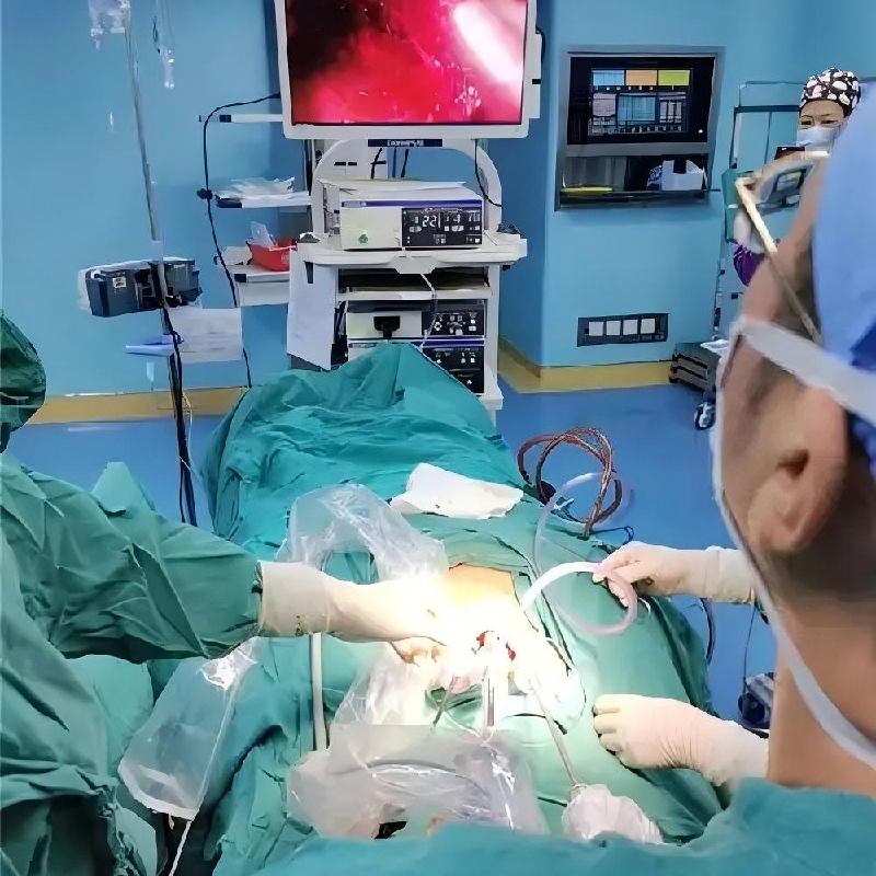
Uzun zamandır, ameliyathanedeki ışıklar sessiz dansçılar gibiydi ve projeksiyon yönleri ve parlaklıkları cerrahın görüşünün netliğini belirliyordu. Titrek mum ışığından göz kamaştırıcı halojen lambalara, verimli LED'lere kadar cerrahi aydınlatma uzun bir evrim geçirdi. Bununla birlikte, tepe ışık kaynaklarının doğal sınırlamaları - kaçınılmaz gölgeler, sık ayarlama gereksinimleri ve derin boşlukların yetersiz aydınlatılması - her zaman cerrahi hassasiyet operasyonlarının "Aşil topuğu" olmuştur. Boşluk aydınlatmasının yükselişi, cerrahi aydınlatmanın "dış projeksiyondan" "iç aydınlatmaya" yeni bir çağını işaret ediyor ve bu sorunlu noktalara devrim niteliğinde çözümler getiriyor.
1. Temel atılım: gölgeleri tamamen ortadan kaldırın ve gölgesiz hassasiyet elde edin
- Boşluk aydınlatmasının temel avantajı, ışık kaynağının doğrudan cerrahi alana ulaşması ve geleneksel dış ışık kaynağının aletlerin, ellerin veya dokuların tıkanması nedeniyle neden olduğu gölgeleri temelden ortadan kaldırmasıdır.
- Geleneksel cerrahide, tavan lambasını ayarlama sıklığı her 7,5 dakikada bir kadar yüksek olabilir (Cerrahi Dergisi'nin araştırma incelemesi), bu da cerrahinin ritmini ciddi şekilde etkiler. İç boşluk ışık kaynakları (retraktörlere, endoskopilere veya özel aletlere entegre edilmiş olanlar gibi) doğrudan hedef alanı aydınlatır ve düzgün, kör açı içermeyen bir ışık alanı sağlar. Amerikan Ameliyathane Hemşireleri Birliği (AORN), cerrahi ortamı optimize etme konusundaki yönergelerinde, görsel yorgunluğu ve çalışma hatalarını azaltmak için gölgeleri ortadan kaldırmanın gerekli olduğunu vurguladı. İç boşluk aydınlatması, bu talebe nihai cevaptır.
2. Hassas yetkilendirme: ayrıntıları aydınlatın, cerrahi kaliteyi ve verimliliği artırın
- İç boşluk aydınlatması, daha konsantre, kontrol edilebilir ve yüksek renk verme özelliğine sahip ışık sağlayarak, özellikle hassas anatomik yapılarda ve derin ve dar boşluklarda cerrahi alanın görünürlüğünü büyük ölçüde artırır.
- Lighting Research and Technology'deki hakemli bir çalışma, iç boşluk aydınlatmasının daha düşük bir ışık yoğunluğunda daha hassas konumlandırma aydınlatması sağlayabildiğini ve cerrahların görsel stresini önemli ölçüde azalttığını doğruladı. Oftalmoloji alanında, kataraktların fakoemülsifikasyonu veya retina onarım ameliyatı gibi, iç boşluk aydınlatması, cerrahi güvenlik ve etkinliği sağlamanın anahtarı olan lens kapsülleri ve retina delikleri gibi milimetre düzeyindeki yapıları açıkça sunabilir (Amerikan Oftalmoloji Akademisi AAO kaynaklarına bakın). Otolaringoloji sinüs cerrahisinde, dar ve kavisli geçitler de iç boşluk ışık kaynaklarının tanıtımı sayesinde açıkça görülebilir.
3. Minimal invaziv cerrahi itici: küçük kesi, büyük başarılar
- İç boşluk aydınlatması, minimal invaziv cerrahinin (MIS) gelişimi için temel itici güçlerden biridir ve daha küçük kesiler yoluyla karmaşık derin ameliyatlar yapmayı mümkün kılar.
- Geleneksel büyük kesi ameliyatları genellikle yeterli ışık ve çalışma alanı elde etmek için yapılır. İç boşluk aydınlatması, "aydınlatma" için kesiyi genişletmeye zorlanmadan, doğrudan vücut boşluğunun derinliklerine ışık kaynağı sağlar. Üroloji alanında, perkütan nefrolitotomi (PCNL) veya böbrek taşları için üreteroskopi, operasyon için küçük kanallar aracılığıyla böbreğe veya üretere ulaşmak için iç boşluk ışık kaynaklarına dayanır. Gastrointestinal cerrahide laparoskopik cerrahi ve jinekolojide histeroskopi/laparoskopik cerrahi, iç boşluk aydınlatması sayesinde minimal travma ile karmaşık prosedürleri tamamlayabilir.
4. Hastalar için iyi haber: iyileşmeyi hızlandırın ve ağrıyı azaltın
- İç boşluk aydınlatması ile desteklenen hassas operasyon ve minimal invaziv yaklaşım, doğrudan daha az doku hasarı, daha az postoperatif ağrı ve daha hızlı iyileşme anlamına gelir.
- Gereksiz doku traksiyonunu ve hasarını azaltmak, daha düşük inflamatuar yanıt ve ağrı seviyeleri anlamına gelir. Çalışmalar, minimal invaziv cerrahinin genellikle kısa hastanede kalış ve hızlı iyileşme süresi avantajlarına sahip olduğunu göstermiştir (minimal invaziv cerrahinin faydaları üzerine NIH incelemesine bağlantı). İç boşluk aydınlatması, bu hedefe ulaşmak için temel teknik garantidir. Hastalar ameliyattan sonra daha hızlı yataktan kalkabilir, komplikasyon riskini azaltabilir ve en kısa sürede normal hayata dönebilir, bu da tıbbi deneyimi ve yaşam kalitesini önemli ölçüde artırır.
5. Güvenlik yükseltmesi: sıcaklığı ve riski azaltın, doktorları ve hastaları koruyun
- İç boşluk aydınlatması (özellikle LED ışık kaynağı), geleneksel aydınlatmanın (özellikle halojen lambalar) neden olduğu termal hasar ve potansiyel elektriksel iletim tehlikesi riskini etkili bir şekilde azaltır.
- LED teknolojisinin kendisi soğuk ışık kaynağı özelliklerine sahiptir ve çok az ısı üretir. Bu, özellikle ışık kaynağı vücuttaki hassas dokunun yakınına yerleştirildiğinde önemlidir ve termal yanık riskini büyük ölçüde azaltır (FDA'nın tıbbi cihazların termal etkilerinin dikkate alınmasına ilişkin kılavuzuna bakın). Aynı zamanda, fiber optik iç boşluk aydınlatma yöntemi, akımın vücut boşluğuna girme olasılığını tamamen izole ederek elektriksel güvenliği daha da artırır ve hastaları ve sağlık personelini korur.
6. Verimlilik ve sürdürülebilirlik: enerji tasarrufu ve tüketim azaltımı, optimize edilmiş çalışma
- İç boşluk aydınlatma sistemleri (özellikle LED tabanlı olanlar), tıbbi kurumlar için işletme maliyetlerini azaltan ve çevresel ayak izini azaltan önemli enerji verimliliği avantajlarına sahiptir.
- LED ışık kaynakları, geleneksel halojen lambalardan çok daha az enerji tüketir. İç boşluk aydınlatması genellikle yalnızca belirli küçük alanları gerektiğinde aydınlatır, büyük tavan lambaları gibi tüm ameliyat masası alanını aydınlatmak yerine, bu da daha fazla enerji tasarrufu sağlar. Tıp endüstrisinde sürdürülebilir kalkınmaya giderek daha fazla önem verilmesiyle (Practice Greenhealth girişimi gibi), düşük enerjili, uzun ömürlü iç boşluk LED aydınlatması, karbon emisyonlarını ve işletme giderlerini azaltmak için akıllıca bir seçim haline geldi.
Çok uzmanlık uygulamaları: tıbbın tüm alanlarını aydınlatmak
İç boşluk aydınlatmasının devrim niteliğindeki değeri birçok tıp uzmanlığına fayda sağlamıştır:
- Endoskopi/tanı: Gastrointestinal sistemi, solunum yolunu ve idrar yolunu aydınlatır, yüksek çözünürlüklü görüntüleme sağlar ve erken kanser taramasına yardımcı olur (kolonoskopi, bronkoskopi gibi).
- Oftalmoloji: Katarakt ve vitreoretinal cerrahi için kritik intraoküler gölgesiz aydınlatma sağlar.
- Otolaringoloji (KBB): Sinüsler, boğaz ve kulak kanalları gibi dar derin alanları aydınlatır (endoskopik burun ameliyatı, laringoskopi gibi).
- Üroloji: Sistoskopi, transüretral cerrahi, nefroskopik cerrahi vb. için uygulanır.
- Jinekoloji: Laparoskopik rahim ameliyatı, histeroskopi ve ameliyatı sağlar.
- Kardiyotorsik cerrahi/girişimsel kardiyoloji: Minimal invaziv kalp cerrahisinde (torakoskopik destekli cerrahi gibi) ve bazı transkateter girişimlerinde derin aydınlatma sağlar.
Geleceğin ışığı: zeka ve özelleştirme
İç boşluk aydınlatmasının evrimi durmaktan çok uzak ve gelecekteki trendler heyecan verici:
- Ayarlanabilir spektrum/renk sıcaklığı: Farklı dokular belirli ışık dalgaları altında daha iyi görselleştirilir ve ayarlanabilir ışık kaynakları doku tanımlamayı ve cerrahi hassasiyeti artıracaktır.
- Kablosuz/uzaktan kumanda: Kabloların kısıtlamalarından kurtulun ve özellikle derin ve karmaşık operasyonlar için cerrahi esnekliği artırın.
- Ultra minyatürleştirme ve modülerleştirme: Daha hassas ameliyatların (nöroşirürji ve mikrocerrahi gibi) ihtiyaçlarına uyum sağlayın ve son derece özelleştirilmiş entegrasyon elde edin.
- Akıllı algılama ve görüntüleme füzyonu: Görünür ışığın ötesinde bilgi boyutları sağlamak için aydınlatma ve endoskopik görüntüleme, floresan navigasyon ve diğer teknolojilerin derin entegrasyonu.
- Sürdürülebilir malzemeler: Tıbbi atık yükünü azaltmak için bazı tek kullanımlık bileşenler için çevre dostu ve parçalanabilir malzemeler keşfedin (EPA tıbbi atık yönetimi bilgilerine bağlantı).
İç boşluk aydınlatması ameliyathanedeki daha parlak bir "ampulden" daha fazlasıdır. Cerrahi görsel yeteneklerin devrim niteliğindeki bir uzantısı ve hassas tıp ve minimal invaziv kavramların uygulanması için temel bir teknik köşe taşıdır. Gölgeleri ortadan kaldırarak, görünürlüğü artırarak, minimal invaziv cerrahiyi mümkün kılarak, iyileşmeyi hızlandırarak, güvenliği sağlayarak ve verimliliği optimize ederek, iç boşluk aydınlatması cerrahinin standartlarını ve sınırlarını yeniden tanımlıyor. Göz küresinin derin içinden dolambaçlı sinüs geçitlerine, atan kalbin kenarından hassas sinir pleksusunun kenarına kadar, iç boşluk aydınlatmasının ulaştığı her yer, cerrahların keskin gözlerini ve yetenekli ellerini göstermeleri için bir sahnedir. Akıllı, kablosuz ve özelleştirilmiş dalgaların yükselişiyle birlikte, iç boşluk aydınlatması, cerrahinin daha yüksek hassasiyet, daha az travma ve daha hızlı iyileşmeye doğru gelecekteki yolunu aydınlatan, asla sönmeyen bir "yaşam ışığı" olmaya devam edecektir. "İç boşluktan" gelen bu ışık huzmesini kucaklamak, cerrahi teknolojinin evriminin bir sonraki zirvesini kucaklamak demektir.